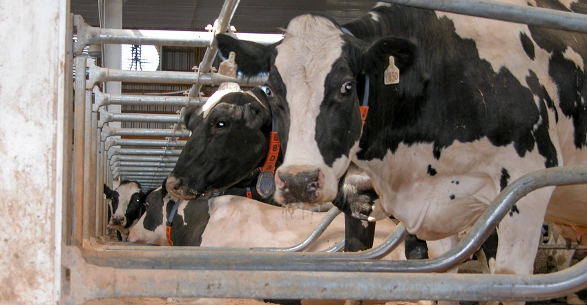
Bovine leukemia virus (BLV) of the family Retroviridae has worldwide distribution and is the cause of enzootic bovine leukosis, a neoplastic disease of lymphatic tissue in cattle.

Bovine leukemia virus (BLV) of the family Retroviridae has worldwide distribution and is the cause of enzootic bovine leukosis, a neoplastic disease of lymphatic tissue in cattle. Most of the infected animals develop a leukemic stage, and approximately 30% progress to persistent lymphocytosis because of polyclonal expansion of B-cells.
This form serves as a reservoir of infection in herds and is deemed a benign stage, whereas 0.1–10% of the infected animals develop tumours, largely B-cell lymphosarcoma. The virus integrates itself into the genome of affected cells as a provirus and is found in the cellular fraction of various body fluids.
Classification and Genomics:
Morphologically, the viral particle, with a diameter ranging between 60 and 125 nm, is constituted by a central electron-dense nucleoid surrounded by an outer viral envelope. Infectious virions contain 60–70 S ribonucleic acids resulting from the association of two 38 S poly-A-containing RNA molecules.
The bovine leukaemia virus envelope gene encodes two disulfide-linked glycoproteins, gp30 and gp5. Glycoprotein 51 is essential for the viral life cycle and plays a critical role in virus-host interaction because it is mainly involved in infectivity.
According to recent research studies based on env gene analysis, BLV has been classified into 10 genotypes (G1–G10) circulating in various geographical regions of the world, with 94.5%–97.7% homology observed among the different isolates of BLV.
In addition to the structural gag, pol, and env genes required for the synthesis of the viral particle, the BLV genome contains an X region located between the envelope and the 3-long terminal repeat, as also observed in other delta retroviruses.
Phylogenetic comparisons of different strains using the pol gene as a reference indicate that BLV and primate T-lymphotropic viruses (PTLV) sequences differ by 42%, thus BLV forms a distinct clade amongst retroviruses.
Within the BLV subgroup, the sequence divergence was below 6% in pol and env, indicating a high degree of conservation among different geographical strains. Although the reasons are unknown, this genomic stability might result from a higher fidelity of reverse transcription or from strict replication constraints.
Clinical Diseases and Pathogenesis:
Clinical Diseases:
Lymphosarcoma in cattle may arise spontaneously (referred to as “sporadic”) or result from infection with bovine leukaemia virus (BLV); the latter is often referred to as an enzootic bovine leukosis. Sporadic lymphosarcoma in cattle is unrelated to infection with BLV. Despite the lack of association, animals with sporadic lymphosarcoma may also be concurrently infected with the virus. Sporadic lymphosarcoma manifests in three main forms:
1. Juvenile lymphosarcoma occurs most often in animals < 6 months old.
2. Thymic lymphosarcoma affects cattle 6–24 months old.
3. Cutaneous lymphosarcoma is most common in cattle 1–3 years old.
Pathogenesis:
The major target of the virus is a B lymphocyte, which expresses surface immunoglobulin M. In addition to B lymphocytes, BLV also persists in cells of the monocyte/macrophage lineage.
Immunoglobulin γ heavy chains are frequently found on lymphoma cells from cattle,consistent with a mature B cell phenotype.
Sequencing of VDJ rearrangements in IgM-secreting B lymphocytes from a BLV-infected cow indicates that IgM antibodies are functional, exhibit polyspecific reactivity, and contain exceptionally long CDR3H. Such long HCDR3s, which are also often found in poor-outcome chronic lymphocytic leukaemia patients (B-CLL), characterise antibodies directed towards negatively charged autoantigens.
Viral infection is followed by a polyclonal expansion of a large and diverse population of lymphocytes harbouring one to five integrated proviruses . At later stages, a few cell clones predominate, and the population evolves towards monoclonality in assays of viral integration.
Proviral integration thus appears to be mandatory for the viral life cycle, although each integration event may not be perfect and, on occasion, some sequences are deleted. In fact, these relatively frequent deletions typically affect the central structural genes and yield dead-end viruses that are unable to replicate in vivo.
The emergence of these deletants might be a fortuitous consequence of viral replication following mistakes during reverse transcription, recombination, or integration.
However, the frequency at which these deletions occur in tumor samples suggests that they provide a selective advantage to the infected cell. In rare cases, it is even possible that a deleted provirus is the sole integrant within the host cell genome.
Transmission:
Cattle are infected with BLV via the transfer of blood and blood products that contain infected lymphocytes. Once infected, cattle develop a lifelong antibody response, primarily to the gp51 envelope protein and the p24 capsid protein. B lymphocytes harbour the integrated provirus but rarely express viral proteins on their cell surface. The exact site of viral replication and expression that drives the immune response remains elusive.
Under experimental conditions, most routes of viral exposure can successfully transmit infection. However, many of these settings are unlikely to be encountered naturally.
Many bodily fluids, including urine, faeces, saliva, respiratory secretions, semen, uterine fluids, and embryos, have been examined for their ability to transmit bovine leukemia virus and are considered to be noninfectious. Only on rare occasions have viruses been found in these fluids.
Colostrum from BLV-positive cows contains viruses and has been found to be infectious experimentally. However, colostrum also contains large amounts of antibody, and it is believed that the protective effects of colostral antibodies outweigh the infectious potential when colostrum is administered in a normal fashion.
Most BLV transmission is horizontal. Close contact between BLV-negative and BLV-positive cattle is thought to be a risk factor. Many common farm practices have been implicated in viral transmission, including tattooing, dehorning, rectal palpation, injections, and blood collection.
Vectors such as tabanids and other large biting flies may also transmit the virus. Vertical transmission may occur transplacentally from an infected dam to the foetus or postpartum from the dam to the calf via ingestion of infected colostrum. Any material that is blood-contaminated or lymphocyte-rich has the potential to infect animals with BLV.
Diagnosis:
- Serologic testing
- Cytology or histologic examination of biopsy samples
Lymphosarcoma is often included on the differential diagnosis list for many diseases because of its wide range of clinical findings.
Viral infection is diagnosed by serology or virology; persistent lymphocytosis is identified by haematology; and neoplastic tumours are identified by histologic examination of biopsies. Positive serology or virology testing for bovine leukemia virus confirms viral infection but not the presence of lymphosarcoma.
Serologic testing by means of ELISA is the most common and reliable way to diagnose infection with BLV. Serology is unreliable in calves that have ingested colostrum from BLV-positive cows because of the passive acquisition of maternal antibodies that typically wane by 4–6 months old.
PCR is a sensitive and specific assay for the diagnosis of bovine leukemia virus infection in peripheral blood lymphocytes. This test can identify the proviral DNA of BLV in the lymphocytes of infected animals and differentiate positive from negative calves in the presence of maternal antibodies.
The diagnosis of lymphosarcoma must be made by cytology or histopathology. Cytologic diagnosis is sometimes difficult because of the frequency of blood contamination in the aspirates.
Control:
- Vaccine
- No vaccine is available.
The most commonly recommended eradication protocol is as follows:
- Identify infected animals using a serologic test
- Cull seropositive animals immediately.
- Retest the herd in 30–60 days.
- Use PCR assays to test young calves and as a complementary test to clarify test results in herds with a low prevalence of infection.
- Repeat testing and culling until the entire herd tests negative.
Testing is then repeated every six months. The herd is declared free when there have been no positive tests for two years. Additions to the herd should have two negative tests 30 and 60 days before arrival.
When test and cull programs are economically untenable, test and segregation programs have been recommended but are rarely implemented. These programs necessitate running two completely separate operations and require additional resources, including money, time, and an available workforce.
Research and Future Perspectives:
Research on the bovine leukaemia virus has been ongoing for several years, with a particular focus on understanding its biology, pathogenesis, and epidemiology. Recent advances in genomic and proteomic technologies have provided new insights into the molecular mechanisms underlying bovine leukaemia virus infections, leading to the development of new diagnostic tools and vaccine candidates.
In the future, it is expected that continued research on the bovine leukaemia virus will lead to the development of effective vaccines, diagnostic tests, and antiviral drugs. Moreover, the knowledge gained from studying these viruses can also have implications for understanding the biology of other retroviruses that affect humans and animals.
Conclusion:
In conclusion, bovine leukaemia viruses are a group of viruses that pose a significant threat to the dairy industry worldwide. These viruses cause a wide range of clinical diseases in animals, leading to substantial economic losses.
However, significant progress has been made in understanding the biology and pathogenesis of bovine leukemia viruses, leading to the development of effective control measures. Continued research on these viruses is necessary to ensure their effective control and to gain insights into the biology of other retroviruses.