Research During The Last Decade Has Made It Increasingly Clear That Autophagy Plays Important Roles In Most Of The Major Human Diseases As Well As In Infection And Immunity,

With increasing evidence for selective autophagy of protein aggregates, organelles and pathogens. This research paper will look into autophagy and how it relates to infection and immunity, specifically in regards to the coronavirus. It will help deduce whether autophagy can be an important factor in ridding the human cells of the virus and becoming a healthy, normally functioning cell again.
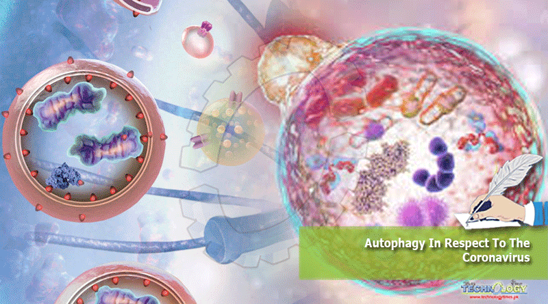
Q. What is autophagy?
Autophagy can be described as a self regulated process, where the damaged, unhealthy cell components are destroyed by the lysosome and removed from the cells. It can be categorised into three subcategories: microautophagy, macroautophagy, and chaperone-mediated autophagy (CMA). In disease, autophagy has been seen as an adaptive response to stress, promoting survival of the cell by removing the non self pathogen, and by breaking down the unneeded proteins in the cells to provide energy. It is known to be the main process that maintains the homeostasis of the cell.
Q. What is Coronavirus?
A virus is a pathogenic, parasitic organism that has no cell membrane, no metabolism, no respiration and cannot replicate outside of a living cell. One such virus is the coronavirus, also known as COVID-19; it is a highly infectious viral disease caused by a strain of the Severe Acute Respiratory Syndrome Coronavirus 2 (SARS-CoV-2). The time from exposure to onset of symptoms is typically around five days but may range from two to fourteen days. The virus is primarily spread between people during close contact, most often via small droplets produced by coughing, sneezing, and talking. Genetic analysis has revealed that the coronavirus genetically clusters with the genus Betacoronavirus , in subgenus Sarbecovirus (lineage B) together with two bat -derived strains.
Q. When and how does autophagy work?
Autophagy is stimulated above the resting rate when nutrients are limited, when cells are under stress, or intracellular bacteria and damaged organelles need to be degraded. The most common initiating factor for autophagy is the lack of basic nutrients in the body, hence a lot of people fast to trigger the process of autophagy in their bodies. Autophagy usually begins after 18-20 hours of fasting. Autophagy is naturally part of the host defence system as it contributes to the discarding of invading microorganisms, which are identified as non-self, by delivering them to the lyso-somal compartment; this is usually referred to as xenophagy . Autophagy works to protect host cells against viral attack by degrading the viruses in autolysosomes. This is done in four steps:
- Initiation : Multitasking protein complex AMPK (adenosine monophosphate-activated protein kinase) is the main protein that signals the initiation of autophagy. AMPK is activated in low energy conditions and it phosphorylates TSC2 and RPTOR to inhibit the mTOR enzyme, which is responsible for cell growth and production along with inhibiting autophagy.This activates the ULK1 enzyme complex, which is responsible for the initiation of autophagy
- Elongation : a phagophore double membrane identifies the antigens of the pathogens and starts extending to engulf the non self pathogens/microorganisms within the cell
- Maturation : the vesicle that had started forming during elongation graduallygrows/extends to become a fully enclosed vesicle with the microorganisms/organelles within it. This vesicle is now known as an autophagosome
- Fusion : completed autophagosomes are then transported through the cell to the lysosomes present to allow the two to fuse together, forming an autolysosome
- Degradation : hydrolytic enzymes (lysozymes/lysosomal hydrolase) present within the lysosome now form enzyme-substrate complexes with the pathogenmolecules (substrates) brought in by the autophagosome, and breakdown the nonself molecules The products are then recycled back to cytoplasm by lysosomal permease
Q. How do viruses affect cells?
Viruses have a self-replicating molecule of RNA or DNA that acts as their genetic code. When they enter the host cell by puncturing the cell membrane, they go straight to the nucleus where transcription for protein synthesis occurs based on the host cell’s DNA. The virus replaces the host’s DNA with its own molecule of DNA/RNA, which causes the mRNA strand transcribed to be based on the virus’s genetic code. The proteins synthesised from this allow the virus to replicate and grow within the host body.
Q. How does the coronavirus enter the host cells and which organs are affected by it?
The virus accesses host cells via the enzyme angiotensin-converting enzyme 2 (ACE2) which is responsible for blood pressure regulation. The virus uses a special surface glycoprotein called a “spike” ( peplomer ) to connect to ACE2 and enter the host cell. The density of ACE2 in each tissue correlates with the severity of the disease in that tissue.
The lungs are the organs most affected by COVID‑19 as ACE2 is most abundantly found in type II alveolar cells , which are only present in the lungs. Other organs affected by the virus include the brain(stem), heart and kidneys.
Q. How does autophagy counter viral infections?
Autophagy controls viral infections at multiple levels by causing the destruction of viruses, promoting antigen presentation and regulating inflammatory responses. This is done in the steps mentioned as part of the process of autophagy.
- The destruction of viruses is done by engulfing them and forming autolysosomes where they are degraded by the lysosomal hydrolase enzymes.
- Following degradation, autophagy coordinates adaptive immunity by delivering virus-derived antigens for presentation to T lymphocytes. The T lymphocytes then have 3 types of cells: T killer cell, T helper cell, and T memory cell. The killer cells directly kill the virus by releasing perforin and cytotoxins. Perforin first makes a pore, or hole, in the membrane of the infected cell. Cytotoxins go directly inside the cell through this pore, destroying it and any viruses inside; The helper cells secrete cytokines to activate B lymphocytes and other T cells; and the memory cells memorise the specific antigen of the virus so the immune response for the next encounter with that specific virus will be much quicker and stronger the next time.
- The inflammatory response is initiated by loading viral components onto endosomal sensors, which triggers the innate immunity in cells in the form of neutrophils and macrophages.
Conclusion: It seems like a plausible solution for cells to undergo autophagy to remove pathogens and other nonself particles that may have invaded the cells and are causing internal damage (which may be in the form of protein synthesis occurring based on the RNA strand of the virus rather than the cell’s own DNA, as in the case of coronavirus), and consequently triggering an immune response to fight off the infection and discard it from the body. This is supported by a study conducted by the La Jolla Institute for Immunology. They tested the blood plasma samples of recovered corona patients, and the results showed that all of the patients carried helper T cells that recognized the SARS-CoV-2 spike protein , which enables the virus to infiltrate our cells. The samples also harbored helper T cells that react to other SARS-CoV-2 proteins, along with virus-specific killer T cells in 70% of the subjects.
Hence, since autophagy helps remove the virus from the cells, along with causing the immune response to be initiated, it is a beneficial process for human cells to go through in order to fight the
This Article was written by Rameen Sajid Cheema