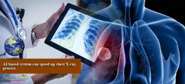
Scientists developed a novel artificial intelligence (AI) system that can reduce the time needed to process abnormal chest X-rays from 11 days to less than three days.

Chest X-rays are routinely performed to diagnose and monitor a wide range of conditions affecting the lungs, heart, bones, and soft tissues, according to the study published in the journal Radiology.
The system reduce the time needed to ensure that abnormal chest X-rays with critical findings will receive an expert radiologist opinion sooner.
The researchers extracted a dataset of half million anonymised adult chest radiographs (X-rays) and developed an AI system for computer vision that can recognize radiological abnormalities in the X-rays in real-time and suggest how quickly these exams should be reported by a radiologist.
The team developed and validated a Natural Language Processing (NLP) algorithm that can read a radiological report, understand the findings mentioned by the reporting radiologist, and automatically infer the priority level of the exam.
By applying this algorithm to the historical exams, the team generated a large volume of training exams that allowed the AI system to understand which visual patterns in X-rays were predictive of their urgency level.
The team, led by Professor Giovanni Montana from the University of Warwick found that normal chest radiographs were detected with a positive predicted value of 73 per cent and a negative predicted value of 99 per cent.
The speedy detection meant that abnormal radiographs with critical findings could be prioritized to receive an expert radiologist opinion much sooner than the usual practice, researchers said.
“Artificial intelligence AI led reporting of imaging could be a valuable tool to improve department workflow and workforce efficiency,” Montana said.
The increasing clinical demands on radiology departments worldwide has challenged current service delivery models, particularly in publicly-funded healthcare systems, researchers said.