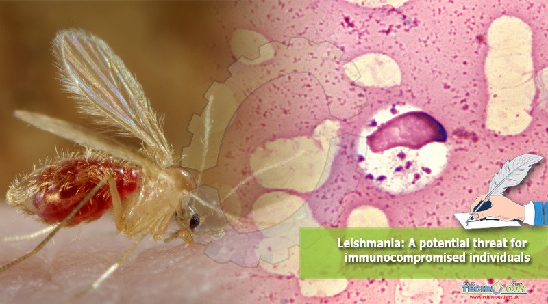
Leishmaniasis is caused by a zoonotically important heamoflagellate, apicomplexan, blood protozone parasite of genus Leishmania.

More than 20 species and subspecies have been characterized on molecular basis. And more than 90 species of sandfly have been isolated to transmit leishmaniasis.
According to a survey conducted by WHO (World Health Organization), 0.7 to 1 million new cases of leishmaniasis occurs annually.
The three clinical forms of disease that have been evident are visceral, cutaneous, and mucocutaneous leishmaniasis.
There are 88 countries on five continents are estimated endemic for leishmaniasis with a conclusion of more than 350 million people are at risk. There are three main forms of disease.
Visceral leishmaniasis
This form also known as kala-azar, it is a serious type of infection whose fatality rate increases upto 95 per cent, if left untreated. It is revealed in different studies that visceral leishmaniasis is caused by three strains of Leishmania donovani.
It is characterized by weight loss, irregular fever and enlargement of visceral organs including liver spleen.
According to an estimation by WHO annually 50,000 to 90,000 population is affected by this disease, throughout the year. The most effected countries include Brazil, India and East Africa.
Post-kala-azar dermal leishmanoid has been observed in most of cases from India, which is characterized by nodules formation all over the body. In case of visceral leishmaniasis, the amastigotes of L. donovani are found in the macrophages of visceral organs.
Cutaneous leishmaniasis
Cutaneous leishmaniasis is a relatively mild type of leishmaniasis and also known as oriental sore.
Cutaneous leishmaniasis is caused by two species including Leishmania mexicana and Leishmania tropica. It is the most common form of leishmaniasis and represents 50-75 per cent of cases.
This disease is characterized by appearance of vascularized papule or nodule on the skin.
Amastigotes of Leishmania mexicana and Leishmania tropica can be found in macrophages around cutaneous sores.
Cutaneous leishmaniasis exists in two forms i.e. (1) the dry or chronic type (2) the moist type or acute type.
It is observed that healing time of this disease increases with secondary infection, while, sores tend to heal within a year in the absence of bacterial secondary infections.
Mucocutaneous leishmaniasis
This disease is caused by Leishmania braziliensis and called as mucocutaneous because of the effected macrophages found in the ulceration area at mucocutaneous junctures of the skin.
The disease is also called as American leishmaniasis, uta, espundia, Pian bois and chiclero ulcer. This is characterized by typical red papules on the skin as primary lesion. These papules ulcerate in one to four weeks and heal in 6-15 months.
Epidemiology
The common sandfly vectors for the transmission of leishmaniasis are phlebotomus major, P. pernicious, P. longicuspis and P. chinensis.
Leishmaniasis exists throughout Mediterranean Basin and extends through South Russia to China in the form of Kala-azar. While the classical form of Kala-azar exists in India and Bangladesh. However, the vector of this form of Kala-azar is Phlebotomus argentipes.
Similarly, most virulent form of Leishmania donovani is transmitted by Phlebotomus spp. in Africa. On the other hand, cutaneous leishmaniasis was recorded endemic in Northern Africa, Europe and Asian Countries including Syria, Israel, Southern Russia, India, China and Vietnam. The prevalence was also observed in Peru, Bolivia, Guianas, Mexico and Brazil.
Mucocutaneous leishmaniasis is also called as American leishmaniasis as this disease is more common in Western Hemisphere from Mexico South to Argentina. Moreover, this disease was found in Sudan, Italy, Kenya, India and China.
In contrast to other forms of leishmaniasis, the sandfly of genus Lutzomyia acts as a vector. The secondary lesion depends on the geographical location. As in Mexico and Central America, the secondary lesion usually appears on ears and called as chiclero ulcer.
Moreover, all three forms of leishmaniasis have zoonotic importance and various forest rodents, cats, dogs and kinkajous act as reservoirs. Additionally, in a study, 15 out of 20 suspected samples were screened positive for Leishmania tropica through nested PCR in 1998. The samples were collected from refugee camp in Pakistan.
Pathophysiology
Leishmania has a complex life cycle involving two hosts a mammal and an invertebrate host as many species of sandflies are involve in transmission of the disease.
As an infected sandfly bites a mammal, the promastigotes are injected under the skin. These promastigotes are ingested by the macrophages and parasitophorus vacuoles are formed.
In macrophages the promastigotes are converted to amastigotes inside the parasitophorus vacuoles. This amestigote undergoes binary fission and large number of amastigotes formed and leads to death of macrophages, which lead to bursting and infection of other macrophages.
A large number of replacement of macrophages leads to enlargement of the visceral organs linked with the reticuloendothelial system including spleen and liver. That decrease in red and white blood cells leads to anemia and leucopenia respectively, secondary bacterial infections and fatality.
However, the infection is restricted to skin tissues in case of cutaneous leishmaniasis and mucocutaneous leishmaniasis.
Interaction with immune competent patients
The leishmaniasis is an opportunistic disease as it mostly effects the children and old population.
According to the WHO, the adult visceral leishmaniasis is an AIDS – related opportunistic disease due to immunosuppression.
The Leishmania spp. resides inside the macrophages. The production of macrophages depends on the activation of CD4 the Th1 cells through release of IFN-ℽ.
As the Th1 cells are under infection of HIV (Human Immuno deficiency Virus), there is a deficiency in Th1 helper cell, moreover, the Leishmnia spp. exposure increases the number of Th2 cells.
Hence, the co-infection of HIV and Leishmania spp. leads to imbalance of Th1/Th2 balance. This imbalance leads to the dysfunction of macrophages activation allowing the Leishmania spp. infection to further exacerbate.
Chemotherapy
In case of visceral leishmaniasis, more extensive chemotherapy and proper nursing is essential. Frequent blood transfusion is a basic necessity in case of more acute visceral leishmaniasis.
In case of visceral leishmaniasis, the intravenous and intra-mascular injections of pentavalent antimony compounds such as antimony sodium gluconate are used with great care. Moreover, lipid-encapsulated Amphotericin B has successfully treated the leishmaniasis.
However, there is no need of treatment in case of cutaneous leishmaniasis which prolongs healing process and increases the rate of secondary infection. So, injections of pentavalent antimony compounds must be used once in a week to decrease the healing time.
Paromomycine can be used tropically to avoid the secondary infection. Similarly, the prolong use of injections of pentavalent antimony compounds is necessary in case of mucocutaneous leishmaniasis.
This article is jointly written by Arsalan Zafar, Muhammad Kasib Khan, Zaheer Abbas, and Hammad Ur Rehman Bajwa from Department of Parasitology, University of Agriculture, Faisalabad.