Matitis in dairy animals: Pakistan is an agriculture based country. The agriculture sector accounts for 20.9 percent of GDP in Pakistan. The agriculture sector has four sub-sectors including crops, livestock, fisheries and forestry.
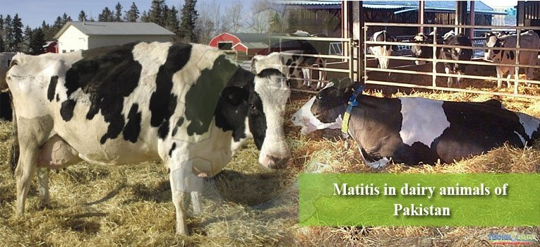
Livestock contains 4.1% in GDP of Pakistan from 20.9 percent of Agriculture. Pakistan is a country producing 5 largest milk yield in the world. It produces about 35.6 billion liters (natural contents) of milk per year.
The most common disease which causes losses in milk production of dairy animals is mastitis It is recorded that about 25 % of the Milk production causes due to mastitis. It causes loses of 240 Million per year.
Mastitis is the inflammation of the mammary gland in the udder, typically due to bacterial infection via a damaged teat. It is also known as lactation mastitis. It is one of the most dangerous disease in dairy animals.
Types of Mastitis
There are several type of mastitis which are discussed below in the table;
|
Acute clinical |
Inflammation of the teat, fever above 39 , weak and dejected animal, lack of appetite. Drastic drop in milk yield. Often follows calving and, less seriously, after cow goes dry. |
|
Hyperacute clinical |
Swollen, red, painful quarter. Milk passes with difficulty. Fever over 41 . Cow has no appetite, shivers and loses weight quickly. Lactation often stops. |
|
Subacute clinical |
No apparent change in udder, presence of flaky particles in milk, especially in initial ejection. Subject appears healthy. |
|
Subclinical |
No symptoms. 15 to 40 cases for every clinical case. Milk appears normal. Only change is detection of pathogenic agent in analysis and increased somatic cell count. Mostly caused by Staphylococcus aureus. |
|
Chronic |
Repeated but mild clinical attacks, generally without fever. Lumpy milk, quarters sometimes swollen. Quarter may become hard (fibrous indurations). Antibiotic treatments often do not work. |
|
Gangrenous |
Affected quarter is blue and cold to the touch. Progressive discoloration from the tip to the top. Necrotic parts drop off. Cow often dies. |
|
Contagious |
Mastitis caused by bacteria such as Staphylococcus aureus and Streptococcus agalactiae, of which other infected cows are the main source. |
|
Environmental |
Mastitis caused by bacteria such as coliforms (e.g. E. coli), of which the main source is a contaminated environment, i.e. manure. |
Economical losses
Mastitis treatment and control is one of the largest costs to the dairy industry in the UK, and is also a significant factor in dairy cow welfare. Losses causes from the following practices.
- Milk thrown
- Thorn away the milk due to contamination by medication or being not fit to drink.
- Reduction in milk yields due to any permanent damage to udder tissues
- Extra workers required for taking care of mastitis’s cows
- Veterinary cares and medicine costs increases.
- Cost of reduced longevity due to premature culling.
Diagnosis of Mastitis
There are different methods to diagnose the mastitis in dairy animals but we are just discussing the most common tests in Pakistan
Direct Microscopic method
- Put 0.1 ml of milk sample on slide
- Dry it and stain it with Newman Lampert’s Stain
- tiCount somatic cells with the help of microscope in certain area
- Multiply the cell counted with a working factor of microscope
- it will give the number of cells per ml of milk.
California Mastitis Test
whenever mastitis occurs, there will be destruction of leukocytes due to phagocytosis. As a result of DNA content increases in milk which is acidic in nature and causes the increase in the acidity of milk. Any alkaline reagent if added, it will neutralize the milk.
A reagent is used in California Mastitis Test which is alkaline in nature. The reagent added in California mastits has alkyl aryl sulfoxide which will cause the precipitation or gel formation in milk.
Surf Field Mastitis Test
This is simplest test which is also used for testation of mastitis
- Make 3 % surf field solution
- Add 6 teaspoons of surf (Washing Powder) in half litre of water
- Shake it to make a good solution
- Filter the solution and heat it.
- Take milk and add equal volume of 3% solution
- Swirl this mixture for half minute
- Examine for precipitation or gel formation (In case of mastitis).
- The test solution is stable for 6 months at room temperature. The solution should be shaken well before use.
Strip Cup Method
It is the simplest method. Take few streaks in cups with black background and observe any abnormality e.g. clots.
Ground Test
Take few streaks on ground. If the absorbance of streak is quick in ground then animal is –ve for mastitis but if the absorbance is slow then milk is mastitic. Late absorbance is due to pus as mastitis milk is pus containing milk.
Microbial culture
|
S aureus |
3% |
|
E Coli |
21% |
|
Yeast |
3% |
|
Enterobacter |
3% |
|
CNS |
5% |
|
Contaminated |
6% |
|
Mixed results |
7% |
|
12 Different Genus |
8% |
|
S environment |
11% |
|
No Growth |
26% |
|
Klebsiella spp |
7% |
Control of Mastitis
There are two main objectives to control the mastitis
- Prevention of new infection in the herd
- Reduction of duration of existing infection
These are the five plans which are most used in mastitis control
- Pre-milking teat dipping
- Post milking teat dipping
- Dry cow therapy
- Prompt treatment of clinical cases
- Culling of chronic mastitic animals from the herd
Treatment
- First you should wash your hands with soap and water
- Wash teats and udder in sanitizing solution
- Thoroughly dry teats and udder with single service individual paper towels
- Dip teats in an effective germicidal teat dip
- Allow 30 seconds of contact time before wiping off teat dip with an individual towel
- Thoroughly scrub the teat end with a cotton swab soaked in alcohol.
- Preferably use commercial antibiotic products in single dose containers designed with partial insertion arrangement formulated for dry cow therapy in single dose containers.
- Do not allow the sterile cannula to touch anything prior to infusion.
- After infusion, remove cannula, squeeze teat end with one hand, massage antibiotic up into the quarter with the other hand.
- Dip teats in an effective germicidal teat dip after treatment.
- One can also prepare infusion solutions and infused with the help of plastic part of IV catheter