Dirofilariasis is one of the zoonotic parasitic infections caused by a filarial worm (nematode), Dirofilaria immitis, primarily affects the canine species (Dog), and is transmitted through the bite of a mosquito, also known as a mosquito-borne filarial worm. The clinical signs and symptoms of dirofilariasis include anorexia, fever, cough, and effusion of the pleural membrane. The treatment includes surgical amputation of lung nodules and granulomas present subcutaneously. Dirofilariasis can also give rise to pulmonary artery granulomas obstructing the pulmonary artery.
By Maria Kausar,Muhammad Sohail Sajid,Kashif Hussain
- Prologue
Dirofilaria immitis belongs to the genus Dirofilaria family Onchocercidae and includes vector-borne filarial pathogens. The most commonly zoonotic reported species are Dirofilaria immitis, D. repens, and D. tenuis. Dirofilariasis in humans is divided into two categories; subcutaneous dirofilariasis (D. repens, and D. tenuis), and pulmonary dirofilariasis (D. immitis).
- Hosts
The definitive hosts for Dirofilaria immitis include dogs, wolves, etc. Mature nematodes can also infrequently be seen in other species such as wild and domestic cats. Mosquito vectors from several genera (Anopheles, Aedes, Culex, Mansonia) play role in the transmission of Dirofilaria species. Definitive hosts for D. repens are wild and domestic canid species and rarely include felids. The only naturally occurring host for D. tenuis is a raccoon and both of these worms are transmitted by a mosquito (Culex, Anopheles, Aedes). D. ursi, the parasite of black bears, is spread by black flies and causes subcutaneous dirofilariasis in bears.
- Signs and Symptoms
The most common symptoms of Dirofilariasis include anorexia, coughing, intolerance to exercise and physical activity, failure to body growth, difficult breathing, discoloration (blue or purple) of the gums and other parts of the skin. Expectoration of blood from the mouth, collapse, bleeding from the nose, and fluid accumulation in the body cavity (Ascites), are the most important signs of Dirofilariasis.
- Pathogenesis
The adult worms of Dirofilaria reside in the right ventricle of the dog’s heart and pulmonary artery. The worms are 12-31 cm long forming aggregation forming an aggregation of almost 50 or more individuals. If aggregations become large, the infection extends to the right atrium, and a load of parasites in the heart increases. Opposing to the belief of canine heartworm disease is not only the consequence of worm load in the heart (ventricle) offering resistance to the flow of blood. Rather, the heart attack in dogs is caused by the deleterious changes in the heart endocardium and pulmonary arteries, resulting in high blood pressure, and hypertrophic ventricle. These pathologies result in reduced cardiac output, lungs infection, difficult breathing, lethargy, prolonged coughing, and ultimately congestive heart failure. Death occurs if proper and urgent treatment is not offered to the dog.
- Signs & Symptoms
The severity of the filarial signs is related to a load of worms present in the lungs and the activity of the dog. The dogs having higher physical activity such as hunters and race dogs will characteristically show more signs of disease as compared to the household and less active dogs. No doubt sedentary dogs have a large number of worms in them but they seldomly show any sign or no sign at all. However, the clinical signs of dirofilariasis can be confirmed by the dead or fragments of the worms in the blood causing blockage of the blood vessels.
- Diagnosis
Diagnosis of dirofilariasis is made based on tissue examination specifically in the areas where inflammation and obstruction can be felt. Inflamed area in the lung is obtained as a part of diagnostic study, the coin-shaped lesions (small round irregularities) on chest x-rays or from the investigation of subcutaneous tissue having nodules in it.
- Discussion
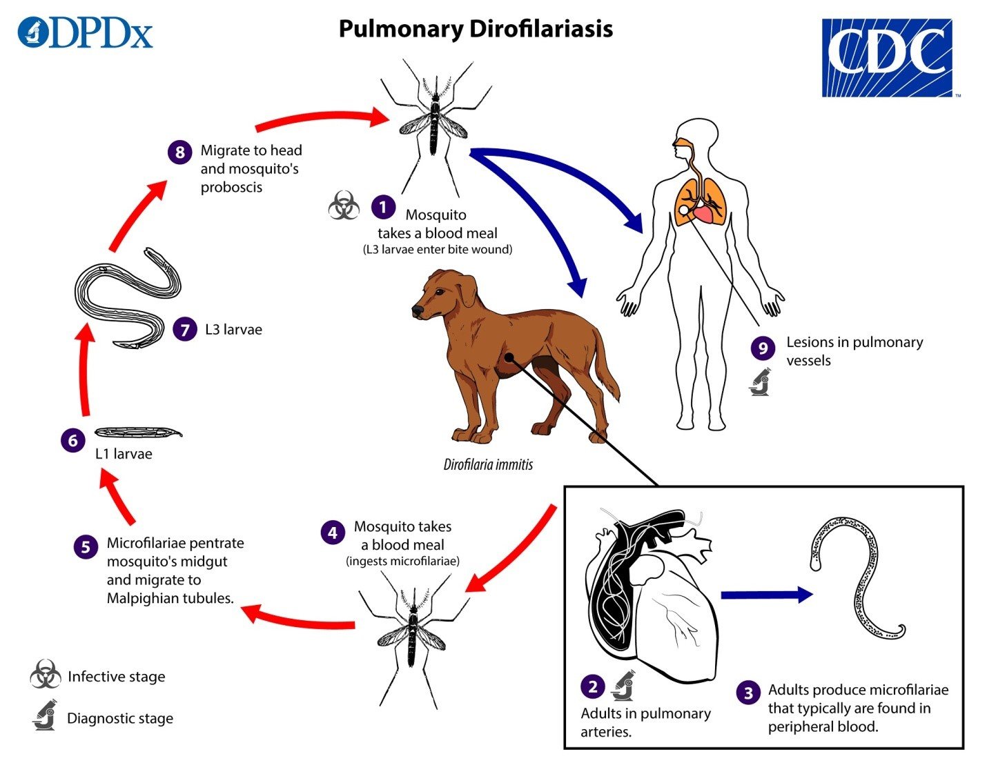
Mosquitoes get infected with Dirofilaria immitis when a mosquito imbibe blood from a dog having microfilaria in it. The mosquito infected with a filarial worm (3rd stage larva) bites its host and penetrates the wound produced by the bite of the mosquito. In the final host, the L3 larvae undergo the process of molting and converting into L4 and sexually mature worms. Adult worm resides in the pulmonary artery and causes infection there. Female nematodes are present in the heart and produce circulating microfilariae present in peripheral blood. The mosquito ingests these microfilariae during a blood meal, microfilariae from the midgut pass through the hemocoel and finally reach the Malpighian tubules and reach in the abdomen. The microfilariae develop into first and third-stage larvae in the Malpighian tubules, from there it migrates to the proboscis of the mosquito and infects another mosquito upon a blood meal.
In humans, Dirofilaria repens represents either a nomadic worm in the subcutaneous tissue or granulomatous nodule, though the reports of pulmonary dirofilariasis with D. tenuis have similar presentation, but may also be found around the conjunctiva of the eye. Due to the conjunctival involvement, the infection in humans is known as Dirofilaria conjunctivae.

- Treatment with precautions
Dirofilariasis infection is preventable. The control of the mosquito population is of prime importance or taking precautionary measures to avoid mosquito bites can reduce the effect of Dirofilariasis in both humans and dogs. Wearing full-sleeve shirts and pants, using mosquito repellents and mosquito nets in the area rich in mosquito population can help in the control of Dirofilaria worm transmission.
Tetracycline therapy has been reported to be effective in damaging D. immitis, even the death of the worm. Prolonged therapy of doxycycline and ivermectin in the replacement of melarsomine injections can help in eliminating the adult worm and decrease the risk of thromboembolism.
Authors:
- Maria Kausar, M.Phil. Parasitology, University of Agriculture, Faisalabad.
- Muhammad Sohail Sajid, Chairman Dept. of Parasitology, University of Agriculture, Faisalabad.
- Kashif Hussain, PhD. Parasitology, University of Agriculture, Faisalabad.