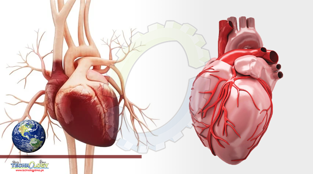
Diagnosis of fetal heart defects, in particular, can improve newborn outcomes and enable further research on in utero therapies, the researchers said.
By Emily Henderson
UC San Francisco researchers have found a way to double doctors’ accuracy in detecting the vast majority of complex fetal heart defects in utero – when interventions could either correct them or greatly improve a child’s chance of survival – by combining routine ultrasound imaging with machine-learning computer tools.
The team, led by UCSF cardiologist Rima Arnaout, MD, trained a group of machine-learning models to mimic the tasks that clinicians follow in diagnosing complex congenital heart disease (CHD). Worldwide, humans detect as few as 30 to 50 percent of these conditions before birth. However, the combination of human-performed ultrasound and machine analysis allowed the researchers to detect 95% of CHD in their test dataset.
Fetal ultrasound screening is universally recommended during the second trimester of pregnancy in the United States and by the World Health Organization. Diagnosis of fetal heart defects, in particular, can improve newborn outcomes and enable further research on in utero therapies, the researchers said.
Typically, the imaging includes five cardiac views that could allow clinicians to diagnosis up to 90 percent of congenital heart disease, but in practice, only about half of those are detected at non-expert centers.
“On the one hand, heart defects are the most common kind of birth defect, and it’s very important to diagnose them before birth,” Arnaout said. “On the other hand, they are still rare enough that detecting them is difficult even for trained clinicians, unless they are highly sub-specialized. And all too often, in clinics and hospitals worldwide, sensitivity and specificity can be quite low.”
The UCSF team, which included fetal cardiologist and senior author Anita Moon-Grady, MD, trained the machine tools to mimic clinicians’ work in three steps.
First, they utilized neural networks to find five views of the heart that are important for diagnosis. Then, they again used neural networks to decide whether each of these views was normal or not. Then, a third algorithm combined the results of the first two steps to give a final result of whether the fetal heart was normal or abnormal.
“We hope this work will revolutionize screening for these birth defects,” said Arnaout, a member of the UCSF Bakar Computational Health Sciences Institute, the UCSF Center for Intelligent Imaging, and a Chan Zuckerberg Biohub Intercampus Research Award Investigator.
“Our goal is to help forge a path toward using machine learning to solve diagnostic challenges for the many diseases where ultrasound is used in screening and diagnosis.”
Originally published at News-medical life sciences